The carotid intima-media thickness test (CIMT) is a non-invasive test that uses ultrasound technology to measure cardiovascular inflammation and plaque.
It is similar to the FIMT (femoral intima-media thickness test). The difference is CIMT probes the carotid artery in the neck, while FIMT probes the femoral artery in the groin.
My CIMT Story
“You’ve got something here,” the ultrasound technician said to me. “We’re technically not allowed to interpret these for you. They have to be read by one of our radiologists, who signs the report.”
I thought he had to be wrong. There was no way I already had plaques. Even though I was already 57 at that time, I believed I lived a healthy lifestyle. I am also a specialist in staying healthy—I trained in and taught preventive medicine at Johns Hopkins.
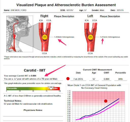
Below is a copy of my first CIMT test done in February 2015. I got an IMT (intima-media thickness) of 0.88 mm. More surprising to me was that I had an arterial age (73) that’s significantly older than me (57).
I mentioned my situation to the technician at a competing CIMT supplier. He wouldn’t exactly confirm, but he said, “I do think you’ll find the report does not show totally clean arterial health.”
That was a kick in the gut. I was frustrated at first. I regretted all the work I’d done, like staying fit and watching my diet. I was also concerned that I had little else I could do.
But a 5-pound weight loss, statins, and aggressive blood sugar management can do wonders with plaque. I actually reversed my arterial age, from 73 years old to 52 years over the next 18 months!
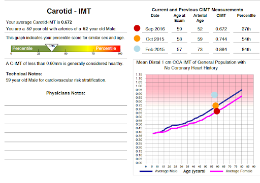
In October 2015, a year after my first CIMT, I was able to reduce my IMT from 0.88 to 0.74. My arterial age was down to 59 years old.
In September 2016, 18 months after my first CIMT, my IMT was further down to 0.67. I had the arteries of a 52-year-old guy.
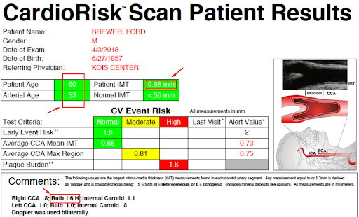
I did my next CIMT in April 2018. This time, I did it with CardioRisk, another CIMT provider. I was 60 years old this time; my arterial age was 53, and my IMT was 0.68 mm. My plaque and arterial age appear to have become stable.
How to Interpret Your CIMT Test Results
Let me explain the things you’ll see in a CIMT report. I’ll use CardioRisk’s report as an example.
Average IMT
This stands for intima-media thickness, the average thickness of the intima-media layer of the carotid artery. In CardioRisk’s report, IMT is labeled as “average CCA IMT.”
IMT is the space between the intima and the media layer. It’s there because of oxidized LDL plaque deposited in that space.
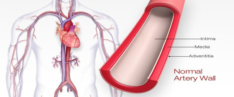
Layers of the Artery. Source: American Stroke Association. Atherosclerosis and Stroke. American Stroke Association website.
Some standards classify average IMT based on the following guidelines:
- less than 0.73mm: normal;
- greater than 0.73mm: abnormal;
- above 0.87 mm: significant inflammation or LDL cholesterol deposition.
There’s no guarantee you’ll get an IMT number with any radiology centers down the street. Be aware that there are other carotid ultrasounds aside from CIMT, and they don’t provide IMT. That’s why you need to ensure that you’re indeed getting a CIMT test.
Arterial Age
The arterial age is estimated based on average IMT.
In the lower-left portion of page 1 of a CIMT report, there’s a graph or “nomogram” which compares your IMT with other people of the same age and gender. You can get your arterial age that would provide a big picture of where you are in terms of the artery aging process.
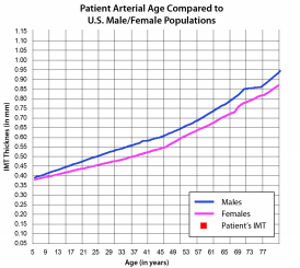
If you got an arterial age that’s higher than your actual age, don’t worry. You can bring your arterial age down with appropriate interventions, like lifestyle, diet, and medications.
Mean Max Average CCA Max Region
Apart from average IMT, mean max is the other important number that confers risk. It is the average (mean) of your largest plaques (max). In CardioRisk’s report, the mean max is labeled as “Average CCA Max Region.”
Think of plaque areas as mountains. Take the top 6 peaks, then average their heights. That’s how the mean max would be calculated.
Mean max is interpreted on the following cut points:
- Less than 0.75mm: normal;
- Between 0.75 to 1.0mm: moderate risk;
- Above 1.0mm: high risk.
Plaque Burden
Plaque burden is the sum thickness of all the discrete plaques. Discrete plaque is any plaque that’s 1.3mm thick or more. Plaque burden is listed in the “Comments” section of a CardioRisk report.
Plaque burden has no cut point. However, “none” means no discrete plaque is detected. Any positive value means there is at least 1 discrete plaque.
For some health professionals, plaque burden is insignificant. However, know that ANY level of plaque means there is cardiovascular disease, as shown by the CAFES-CAVE study.
Published in Atherosclerosis in June 2001, the CAFES-CAVE study looked into 10,000 “healthy” subjects for 10 years. It was done on a population made up of 60% male and 40% female, with ages ranging from 35 to 65 years.
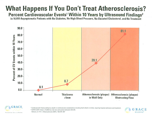
This kind of CIMT study was done to find out about the subjects’ artery conditions. In the graph above, you can see how plaque thickness correlates with the likelihood of getting cardiovascular disease (heart attack or stroke) in the next 10 years.
Types of plaque
Apart from average IMT and mean max, a CIMT test can also provide information on whether the detected plaque is soft or calcified.
In CardioRisk’s reports, plaque is categorized into:
- “S” stands for soft;
- “H” for heterogeneous;
- “E” for echogenic. (Echogenic plaque is a hard or calcified plaque. “Echogenic” means it blocks ultrasonic waves.)
As for me, I’ve retained heterogeneous plaque since my first CIMT. I haven’t been able to get it totally calcified, nor had it totally soft.
Patients come to me, saying they wanted to get rid of all their plaque. While plaque reversal isn’t a simple thing to do, I tell these patients that they can stabilize their plaque.
How? By calcifying it.
Soft plaque vs. Calcified plaque
A study titled “Echolucent carotid plaques predict future coronary events in patients with coronary artery disease” published in JACC (Journal of the American College of Cardiology) in 2004.
This study looks at hard/calcified/echogenic plaque vs. soft/echolucent plaque. (“Echolucent” means letting ultrasonic waves pass through. “Echogenic” bounces ultrasonic waves.)
Here are some images from the study. You’ll see the soft/echolucent on the left and the hard/calcified/echogenic plaque on the right.
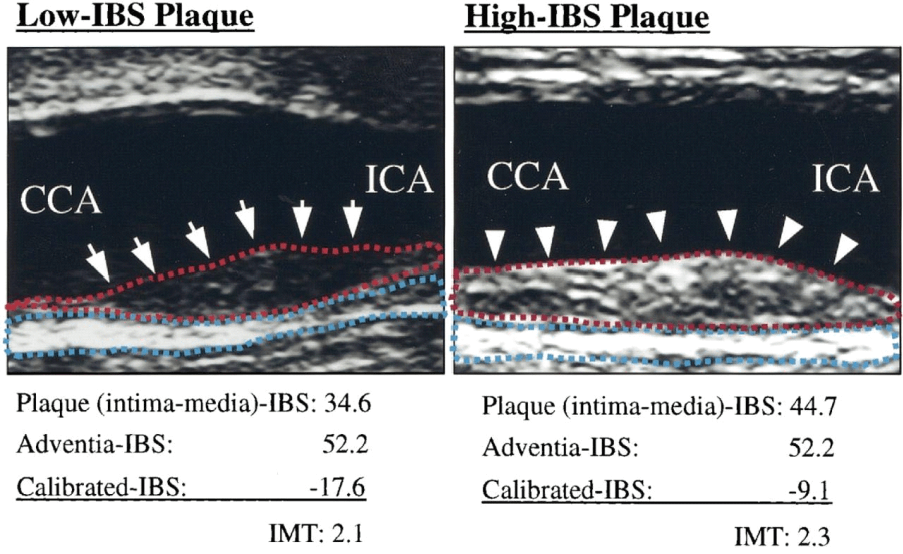
Soft plaque vs. calcified plaque – Appearance. The left shows “low IBS plaque,” which means soft or “hot” plaque, or in this study, “echolucent” plaque. The image on the right shows “high IBS plaque,” also referred to as calcified, hard, or “echogenic” plaque. (Echolucent plaques let ultrasonic waves pass through with no echo; thus, they appear to be transparent. Echogenic plaques reflect ultrasound waves; they appear as solid white mass.) Source: Honda O, Sugiyama S, Kugiyama K, Fukushima H, et al. Echolucent carotid plaques predict future coronary events in patients with coronary artery disease.
How do we know then whether calcified plaque is more stable than soft plaque?
Researchers demonstrated the stability of calcified plaque by using Kaplan-Meier (life table) analysis. The analysis works this way: every time someone with soft plaque has a cardiovascular event, there will be a downtick in the soft/echolucent plaque line. This premise also applies to the hard/calcified/echogenic plaque line.
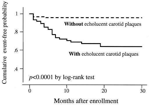
Kaplan-Meier Analysis. Source: Honda O, Sugiyama S, Kugiyama K, Fukushima H, et al. Echolucent carotid plaques predict future coronary events in patients with coronary artery disease.
Here are the findings:
- Out of 215 subjects, 112 had soft/echolucent plaque, while 103 had hard/calcified/echogenic plaque.
- Out of 103 subjects with hard/calcified/echogenic plaque, only 4 had events (3.9%).
- Out of 112 subjects with soft/echolucent plaque, 29 had events (25.9%).
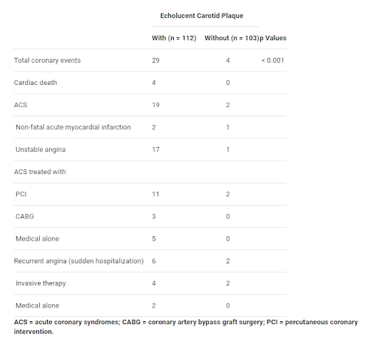
Echolucent vs. Echogenic plaques. Source: Honda O, Sugiyama S, Kugiyama K, Fukushima H, et al. Echolucent carotid plaques predict future coronary events in patients with coronary artery disease.
As these data have revealed, there’s indeed a big difference between having soft/echolucent soft plaque and having hard/calcified/echogenic plaque.
Advantages of CIMT
CIMT is Not Just Any Carotid Ultrasound
While CIMT uses ultrasound, it differs from other carotid ultrasound tests (like the carotid Duplex). Other carotid ultrasound tests usually check only blood flow restriction. CIMT can do more; it can measure inflammation and/or plaque visualized as IMT.
Thus, it’s important to note this difference when visiting a radiology center to be sure you are indeed getting a CIMT test and not another carotid ultrasound test. I suggest you approach only known CIMT providers. (I have recently done mine with CardioRisk).
CIMT Detects Both Soft and Hard Plaques
CIMT is the only plaque screening test that can characterize the type of plaque—whether plaque is soft and dangerous, hard and stable, or a mix of hard and soft. This is CIMT’s primary advantage over the coronary calcium score test.
CIMT vs. Calcium Score & Other Plaque Screening Tests
CIMT uses sound waves. Unlike calcium scans and angiograms, CIMT doesn’t use X-rays. Unlike angiograms, it doesn’t use any contrast dye injected into the patient’s arteries. Unlike cardiac catheterization, it doesn’t involve a catheter that’s inserted into the arm or groin.
Given these advantages, why has CIMT been ignored? The key weakness is that CIMT suffers from a lack of standardization. The quality of the test also depends on the skills of the CIMT technician.
Still, CIMT remains an excellent test in many aspects. I would recommend it if there’s a high-quality CIMT provider near you. And it’s inexpensive, too.
If you found this article helpful and want to start taking steps toward reversing your chronic disease, Dr. Brewer and the PrevMed staff are ready to serve you no matter where you’re located.
To find out more, schedule a consult here: prevmedhealth.com
References
Belcaro G, Nicolaides AN, Ramaswami G, Cesarone MR, et al. Carotid and femoral ultrasound morphology screening and cardiovascular events in low risk subjects: a 10-year follow-up study (the CAFES-CAVE study(1)). Atherosclerosis. 2001 Jun;156(2):379-87. doi: 10.1016/s0021-9150(00)00665-1.
Cedars-Sinai. Carotid Intima-Media Thickness Test (CIMT). Cedars-Sinai website. https://www.cedars-sinai.org/programs/heart/clinical/womens-heart/services/cimt-carotid-intima-media-thickness-test.html. Accessed January 20, 2021.
Chambless LE, Folsom AR, Clegg LX, Sharrett AR, et al. Carotid wall thickness is predictive of incident clinical stroke: the Atherosclerosis Risk in Communities (ARIC) study. Am J Epidemiol. 2000 Mar 1;151(5):478-87. doi: 10.1093/oxfordjournals.aje.a010233.
Chambless LE, Heiss G, Folsom AR, Rosamond W, et al. Association of coronary heart disease incidence with carotid arterial wall thickness and major risk factors: the Atherosclerosis Risk in Communities (ARIC) Study, 1987-1993. Am J Epidemiol. 1997 Sep 15;146(6):483-94. doi: 10.1093/oxfordjournals.aje.a009302.
Cheng HG, Patel BS, Martin SS, Blaha M, et al. Effect of comprehensive cardiovascular disease risk management on longitudinal changes in carotid artery intima-media thickness in a community-based prevention clinic. Arch Med Sci. 2016 Aug 1;12(4):728-35. doi: 10.5114/aoms.2016.60955.
Costanzo P, Perrone-Filardi P, Vassallo E, Paolillo S, et al. Does carotid intima-media thickness regression predict reduction of cardiovascular events? A meta–analysis of 41 randomized trials. J Am Coll Cardiol. 2010 Dec 7;56(24):2006-20. doi: 10.1016/j.jacc.2010.05.059.
Honda O, Sugiyama S, Kugiyama K, Fukushima H, et al. Echolucent carotid plaques predict future coronary events in patients with coronary artery disease. J Am Coll Cardiol. 2004 Apr 7;43(7):1177-84. doi: 10.1016/j.jacc.2003.09.063.
Integrative Medicine Center of Western Colorado (IMC). CIMT ultrasound screening and results. IMC website. https://imcwc.com/cimt-measurements. Accessed January 20, 2021.
Revolution Health & Wellness. How To Read Your CIMT Test Report. Revolution Health & Wellness website. https://www.revolutionhealth.org/how-to-read-your-cimt-test-report. Accessed January 20, 2021.
Stein J. Carotid Intima-Media Thickness Progression and Cardiovascular Disease Risk. J Am Coll Cardiol. 2011 May 31;57(22):2291. https://doi.org/10.1016/j.jacc.2010.12.035.

